


RIRS Laser Kidney Stone Surgery in Indore – What Actually Happens During the Operation?
By Dr. Yusuf Saifee | Consultant Urologist, Laparoscopic & Robotic Surgeon, Kidney Transplant Surgeon
Many patients come to us with kidney stones and have one common concern. Their doctor has advised RIRS or laser surgery, but they are not really sure what happens during the operation.
Will there be any cuts? How does the laser reach the kidney? Will the entire stone come out? And what if some small fragments are left behind?
These are very common questions.
Today, I want to explain how we usually deal with a stone of around 10–15 mm, particularly when it is stuck in the upper ureter or located inside the kidney.
I will explain it in the same way I usually explain things to my patients, step by step.
A 10–15 mm Stone in the Kidney or Ureter
Sometimes patients continue taking medicines for months, hoping that the stone will eventually pass in the urine.
But the size and location of the stone matter a lot.
A 3–4 mm stone and a 10–15 mm stone are completely different situations.
A small stone may pass naturally. But when a 10–15 mm stone is stuck in the ureter, the chances of spontaneous passage are quite low.
And if it is blocking urine drainage, the kidney may gradually become swollen. We call this hydronephrosis.
This is something I always explain to patients. Pain is not the only thing we worry about. We also need to protect kidney function.
On the other hand, a stone lying inside the kidney may not be causing any obstruction or symptoms. Such stones do not always require immediate surgery. We look at the size, location, symptoms, growth and other factors before deciding.
First, We Enter Through the Natural Urinary Passage
Now, how do we actually reach the stone?
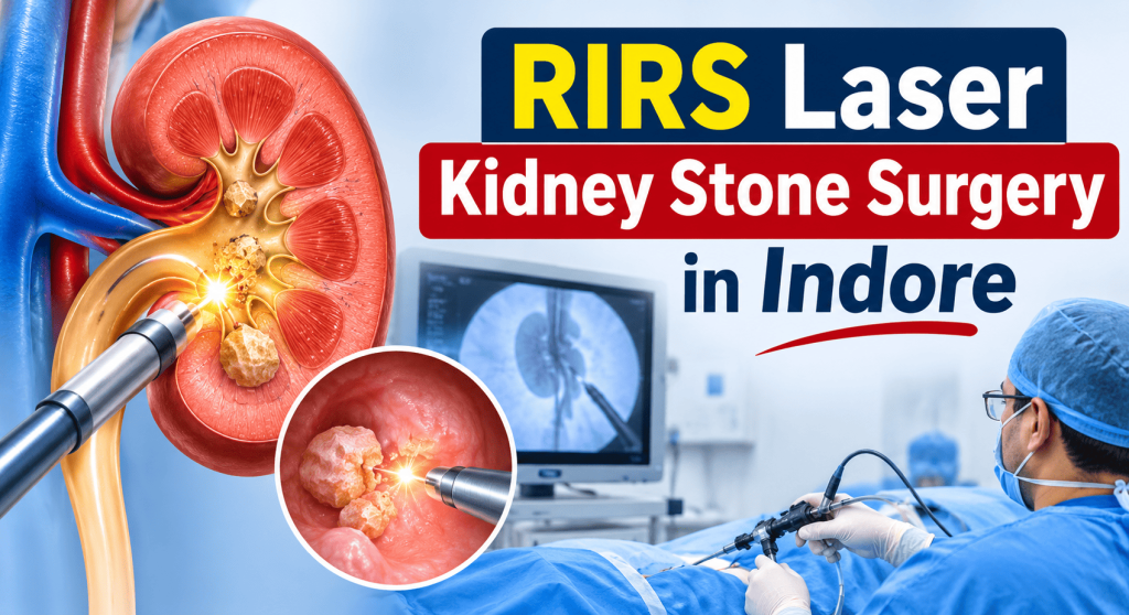
In RIRS or flexible ureteroscopic laser surgery, we don’t need to make a cut on the abdomen.
We pass a very thin camera through the normal urinary passage into the bladder.
Inside the bladder, there are two small openings, one for each ureter.
We identify the opening on the side where the stone is located.
Then, very carefully, we pass our instruments into the ureter.
The important thing is that we are not working blindly. We can see the inside of the urinary passage on a monitor.
When we reach the stone, we assess its position, whether it is impacted, and whether there is swelling around it.
If the stone is inside the kidney, we take our flexible scope further up into the kidney and locate it.
Why Do We Sometimes Push the Stone Back into the Kidney?
This is an interesting part of the procedure.
Suppose a stone is stuck in the upper ureter.
There may be very little space around it, particularly if the stone has been impacted for a long time.
In selected cases, if the stone can be moved safely, we may gently reposition it into the kidney.
Why?
Because inside the kidney, we may have more space to work with the flexible scope and laser.
We can position the stone in a suitable calyx and break it under direct vision.
But this is not necessary or possible in every case. Sometimes we break the stone directly inside the ureter itself.
The decision depends on the location of the stone, the condition of the ureter, and how safely we can access it.
Now Comes the Laser
Once we have reached the stone, we pass a very thin laser fibre through the scope.
The fibre is quite small, but it can deliver enough energy to break a hard stone.
You can actually see the stone breaking on the monitor.
Depending on the stone and the technique, we may break it into small pieces or convert much of it into very fine dust.
This is called laser dusting.
With newer suction-assisted techniques, we can also remove some of the stone dust and fragments during the procedure.
One question patients ask is whether the laser can damage the kidney.
The laser is used under direct vision, and we carefully control where the energy is delivered.
There is still a risk of tissue injury, bleeding, or other complications, as with any endoscopic surgery. That is why proper
